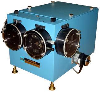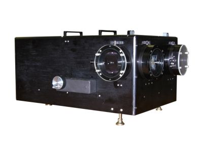
Very High Throughput Monochromator / Spectrometer
- f/3.4 with oversize grating, 3.6 with 120*140mm SNAP IN
- High Efficiency Broadband Al+MgF2 Coatings
- Precision Micrometer Adjustable Slits
- Dual Turret (f/6.9) available as an option
The three versions of the Model 205 have been developed with researchers to meet diverse needs of spectral analysis. The half meter focal length is popular. It provides reasonable spectral resolution, throughput and package size. Every lab has room for a half meter monochromator! Each application is unique and the versions of the McPherson Model 205 will allow you to tailor the instrument to produce better data from a given experiment. Used in air and with a selection of gratings, the spectral range of the 205 extends from 185 nanometersm to 20 microns or more. Multiple ports are available for mounting accessories or CCD detectors, etc. McPherson SNAP IN gratings allow the alignment-free use of many different gratings. All versions work with our digital scan control for scanning or with corrected optics for imaging, all but the quad turret 205wr can be configured for both.
Model 205 PDF Data Sheet
Specifications & Additional Information:
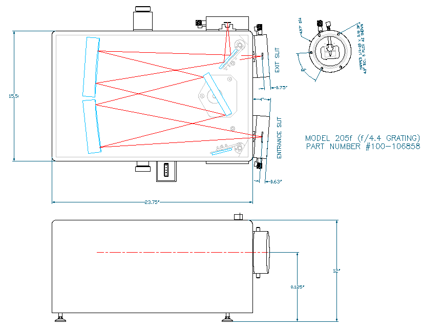
| Optical Design | Czerny Turner with kinematic grating mount |
| Focal Length | 500 mm |
| Aperture Ratio | f/4.3, f/3.2 with single grating holder, f/6.9 with turret |
| Wavelength Range | refer to grating of interest for range |
| Wavelength Accuracy | +/-0.2nm (on counter, with 1200 G/mm grating) |
| Wavelength Reproducibility | +/- 0.01nm (with 1200 G/mm grating) |
| Slit Locations | Axial and lateral with optional extra entrance and exit port selection mirrors depending on version |
| Focal Plane | 25mm, multiply dispersion by the width of your detector for range |
Performance with various diffraction gratings:
| Grating (G/mm) (others available) |
3600 |
2400 |
1800 |
1200 |
600 |
300 |
150 |
75 |
50 |
| Wavelength Range from 185-nm to |
430nm |
650nm |
860nm |
1.3um |
2.6um |
5.2um |
10.4um |
20.8um |
78um |
| Resolution (nm)1 |
0.016 |
0.02 |
0.03 |
0.04 |
0.08 |
0.16 |
0.32 |
0.64 |
1 |
| Dispersion (nm/mm) |
0.5 |
0.83 |
1.1 |
1.66 |
3.33 |
6.7 |
13.3 |
26.6 |
40 |
| First Order Littrow Blaze (nm)2 |
Holo | Holo | Holo | Holo | 300nm | 750nm | 1.25um | 2.0um | 45um |
| | 240nm | | 250nm | 500nm | 1.0um | 2.5um | 3.0um | |
| | 300nm | | 300nm | 750nm | 3.0um | 4.0um | 8.0um | |
| | | | 500nm | 1.0um | 4.0um | 6.0um | 10.0um | |
| | | | 750nm | 1.85um | | 8.0um | 12.0um | |
| | | | 1.0um | | | | | |
1. Tested in scanning mode at 312/313 nanometers with 10 micron wide slits and at slowest aperture ratio
2. Gratings work best from 2/3 blaze wavelength to 3/2 blaze wavelength
Outline Drawing
Select Publications
Abstract: A laser based spectrofluorometer with f/# matched optics has been developed for the investigation of metal ion binding sites on nonviable biomaterials. Solid biomaterial samples have been encapsulated in a KBr glass matrix for these investigations. This sample preparation technique has allowed for the investigation of metal ion binding sites by both luminescence and infrared spectroscopy. The utility of the system and the method of sample preparation has been demonstrated with Eu(III) bound to cultured cell fragments from Datura innoxia.
Lawrence R. Drakea, Trevor R. Bakera, Peter C. Starka & Gary D. Raysona
Abstract: Dissociative excitation of CH3COCN to produce CN(B-X) and CN(A-X) fluorescence was studied by resonance enhanced multiphoton excitation at 292 nm. The laser power dependence of the CN(B-X) fluorescence intensity and the lifetime of the one-photon excited S1 state showed that CN(B) formation takes place in the direct two-photon and two-body dissociation mechanism, CH3COCN+2hν →CH3CO(X̃)+CN(B). Vibrational and rotational energy distributions of the nascent CN(B) fragment were determined by a simulation analysis of the dispersed fluorescencespectrum. The vibrational distribution was found to be of the relaxed type and rotational distribution in each vibrational state could be approximated by a Boltzmann distribution. The best-fit vibrational distribution of CN(B) was Nv′=0: Nv′=1:Nv′=2=1.00: 0.25: 0.07 with the respective rotational temperatures of Tr(v′=0)=2600 K, Tr(v′=1)=1000 K, and Tr(v′=2)=900 K. The internal state distributions were found to be hotter than those predicted by the statistical model with complete energy randomization within the excited molecule. The results indicate a dissociation mechanism where both the vibrational energy deposition in the photoexcitation and available energy redistribution before the bond breakage are limited within the modes of the skeletal CCOCN structure. Possible decay channels other than the CN(B) production, upon two-photon excitation at 292 nm, are also discussed based on the potential surfaces previously predicted. The formation of CN(A) presently observed in the direct two-photon excitation can be interpreted as the dissociation of the electronic excited intermediate states, populated competitively via internal conversion(s) from the upper electronic states. To obtain a deeper understanding of higher excited states of acetyl cyanide, the vacuum UV absorption cross section was also determined in the 110–200 nm region, using a synchrotron radiation source.
Jun-ichi Aoyama, Takashi Sugihara, Kiyohiko Tabayashi and Ko Saito
Abstract: The formation of excited states resulting from electron capture by Auq+ ions (q from 12 to 18, v = 2v0) colliding with H2 has been measured by means of optical methods. The capture cross sections into various n, l states are derived from measured emission cross sections by applying a simplified cascade correction. It is found that high l states are preferentially populated and that a large number of n states contributes to the total capture process. The general picture, which emerges from the present study, is in reasonable agreement with theoretical calculations by Olson, by Ryufuku and Watanabe, and by Crothers.
P Hvelplund, E Samsoe, L H Andersen, H K Haugen and H Knudsen
Abstract: The first excited electronic state of TII was investigated by laser excitation and laser fluorescence spectroscopy. Almost completely resolved rotational fine structure was observed with the help of a collimated molecular beam and a single-mode cw dye laser. From the derived Dunham parameters Ylk a RKR potential for the excited state is calculated. The unusual shape of the potential energy curve can be understood as originating from an avoided crossing of the ionic ground-state potential and a non-bonding covalent state of the atomic asymptote (6p) 2P(TI)+(5p)52P(I).
J. Kieckhäfer, H. Knöckel, E. Tiemann
Abstract: Standoff time-resolved Raman spectroscopy with visible (532 nm) laser excitation is a powerful technique for mineral detection of planetary surfaces (e.g., [1-3]). Some of the minerals, however, show strong fluorescence interference with 532 nm laser excitation (e.g., [4]). It is known that excitation of samples with deep UV could minimize fluorescence interference with the Raman spectra of minerals. In addition, the intensity of the Raman scattering is anticipated to be high for samples excited with UV laser due to 1/λ4 dependence of the scattered light. We have carried out preliminary work to evaluate the feasibility of using 266 nm laser excited Raman spectroscopy for detecting minerals
S. K. Sharma, T. E. Acosta-Maeda, J. N. Porter, A. K. Misra, and S. M. Angel